

MIT Teaches AI About Symmetry to Get Clearer Brain Scans of Babies Moving in the Womb
Researchers from MIT, Boston Children's Hospital and Inria have built E(3)-Pose, a deep learning model that uses the left-right symmetry of the fetal head to track its motion during MRI, so the scanner can adjust and produce higher-quality 3D images for radiologists.
Artificial IntelligenceGNGV Editorial Team4 min readLast reviewed October 26, 2026
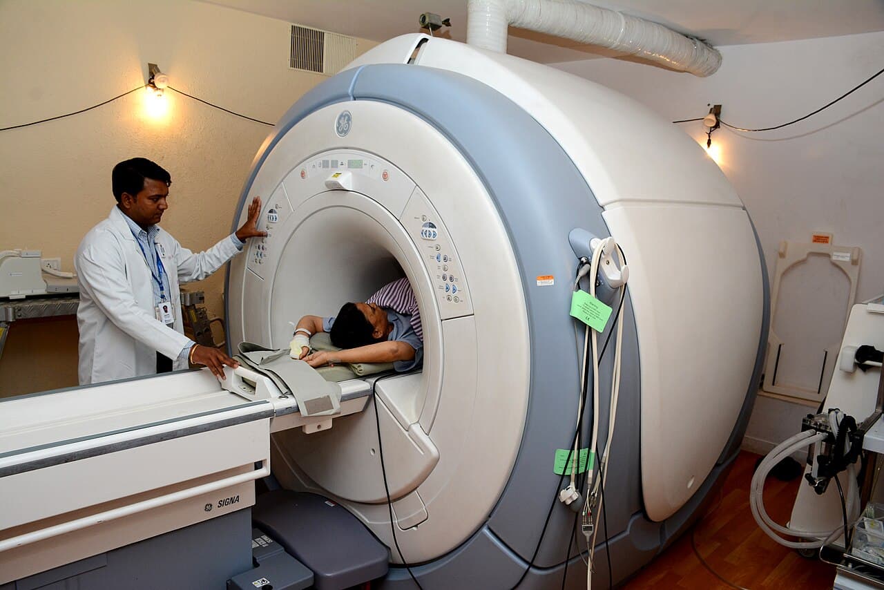
Magnetic resonance imaging can reveal a baby's developing brain in remarkable detail, but there is a catch: babies in the womb move. When a fetus shifts during a scan, the thin two-dimensional slices that make up a 3D image can end up coming from different angles, leaving gaps and odd orientations that make abnormalities easier to miss. Researchers at MIT have developed an AI system designed to keep up.
The model, called E(3)-Pose, was described by MIT News on 19 October 2026 and presented at the 2026 European Conference on Computer Vision. It tracks the motion of the fetal head during an MRI scan so that the scanner can adjust the slices it acquires and produce a higher-resolution 3D scan for radiologists. The team hopes this will reduce missed or incorrect diagnoses of fetal brain abnormalities and spare families repeat hospital visits.
The key idea is to build knowledge of the body into the AI rather than making it learn everything from data. E(3)-Pose embeds the left-right symmetry of the fetal head into its architecture, which helps it work out the brain's orientation even when slices arrive at unusual angles. "One of the problems with having the baby move is that the slices are all over the place," said first author Ramya Muthukrishnan, an MIT PhD student in the lab of Professor Polina Golland. "Ramya's work elegantly combines geometric concepts like equivariance and symmetry with the power of deep learning," said Golland, one of the senior authors.
The project brought together MIT, Boston Children's Hospital and the French research institute Inria. Borjan Gagoski of Boston Children's and Harvard Medical School developed the imaging sequence used to acquire additional scans for training, and research assistant Aryn Lee recruited pregnant volunteers. Senior author Ellen Grant, who directs the Fetal Neonatal Neuroimaging and Developmental Science Center at Boston Children's, said the approach could help automate fetal brain MRI, "replacing today's slow, manual process with faster and more comfortable scans for mothers."
This is research, not yet routine care. MIT News reports no headline accuracy figures, the model was trained mainly on third-trimester fetuses, and getting it to run on the scanner was difficult, with repeated crashes during development. The team's next goal is to handle younger fetuses, which move even more. If that succeeds, expectant parents could one day face fewer repeat scans and clearer answers.
Why it matters
Sharper, more reliable fetal brain scans could mean fewer missed diagnoses and fewer stressful repeat appointments for expectant families.
Sources
What you can do
Read the full MIT News article to learn how the system works; for questions about prenatal imaging, talk to your own care team.


